Fig. 2.
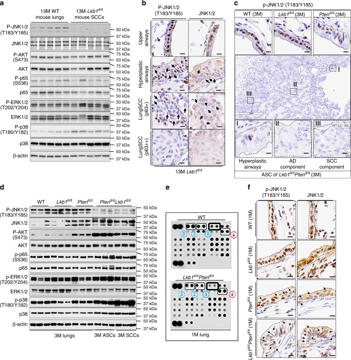
p-JNK1/2 are reduced in Lkb1-deficiency-driven mouse LSCC development. a Western blot (WB) analysis (n = 5) of protein expression in 13-month-old (13M) Lkb1d/d mice. b Representative P-JNK1/2 and JNK1/2 IHC staining of mouse lungs (n = 6). Dashes outline regions of interest. P-JNK1/2 staining is usually nuclear while JNK1/2 staining is usually cytoplasmic and nuclear. c Representative IHC staining results of P-JNK1/2 in 3-month-old mouse lungs and lung tumors (n = 6). Black dashes point to the areas of interest. Box I: hyperplastic airway; Box II: adenocarcinoma (AD) component; Box III: peripheral squamous cell carcinoma (SCC) component. Scale bar: 300 μm (black box) and 10 μm (dark bar). d WB analysis of 3-month-old (3M) mouse lungs and lung tumors. e Kinome array was performed in 1-month-old (1M) mouse lungs (n = 6) (see Supplementary Fig. 4b for a full summary of kinome array). The dots in the membranes are anti-phosphorylated (P)-proteins and the numbers pointing out the top changed proteins were listed in Table 3. f Representative IHC staining of P-JNK1/2 and JNK1/2 in 1-month-old mouse lungs (n = 5). Arrows point to the cells with weak nuclear staining. Scale bar: 300 μm (Black box) and 10 μm (dark bar)

