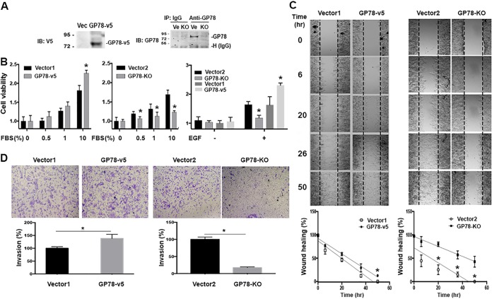
FIG 4.
GP78 promotes cancer cell proliferation, motility, and invasion. (A) Western blot analyses confirmed GP78 overexpression (left) and GP78 knockout (right) in Huh7 cells. Numbers to the left of the gels are kilodaltons. (B) MTT assays of GP78-overexpressing (GP78-v5), overexpressing vector control (Vector1) GP78 knockout (GP78 KO), and knockout control (Vector2) Huh7 cells. Cells were seeded overnight and then cultured in medium containing FBS or EGF (100 ng/ml), and cell proliferation was measured after 24 h. The quantitative data are presented as means and SD from the results of 3 independent experiments (*, P < 0.05). (C) Wound-healing assays of the indicated Huh7 cells cultured in serum-free medium in the presence or absence of EGF for the indicated times. Of note, EGF without FBS was sufficient to promote cell migration. The graphs were generated after normalization with the average distance of the scratched area (n = 3; *, P < 0.05). (D) (Top) Migration and invasion assays of the indicated Huh7 cells incubated with 1% FBS plus 100 ng/ml EGF for 24 h using Transwell chambers. (Bottom) Numbers of cells migrating through the Transwell membrane. The quantitative data are presented as means ± SD from the results of 3 independent experiments (*, P < 0.05).

