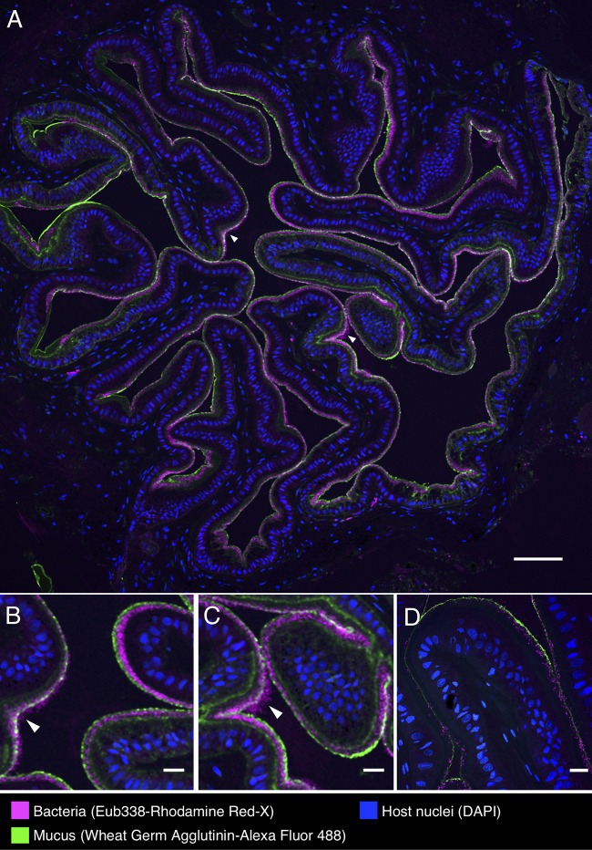
FIG 5.
Spatial organization of bacteria in the esophagus of the European common cuttlefish, S. officinalis. The images shown are cross-sections of esophagus that were embedded in methacrylate, sectioned, and subjected to fluorescence in situ hybridization (FISH) with near-universal bacteria probe (Eub338) and fluorophore-labeled wheat germ agglutinin to visualize mucus. (A) Bacteria (magenta) lining the interior of the esophagus in a control animal in association with the mucus layer (green). Host nuclei (DAPI staining) are shown in blue. Panels B and C are enlarged images of the regions marked with arrowheads in panel A where bacteria extend past the edge of the mucus layer. (D) Esophagus from an antibiotic-treated animal. Scale bars = 100 μm in panel A and 20 μm in panels B, C, and D.

