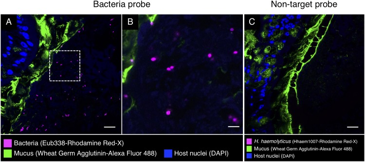
FIG 7.
Fluorescence in situ hybridization in intestine of S. officinalis. Shown is a methacrylate-embedded section of a pilot investigation animal hybridized with the near-universal probe Eub338 and stained with fluorophore-labeled wheat germ agglutinin to visualize mucus. (A) Bacteria (magenta) are sparsely distributed through the lumen. (B) Enlarged image of the dashed square in panel A. (C) An independent FISH control with a nontarget probe (Hhaem1007). No signal was detected. Scale bars = 20 μm in panels A and C and 5 μm in panel B.

