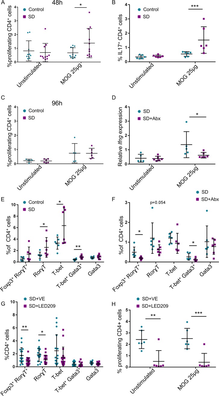
FIG 4.
Microbiota-dependent autoreactive response of the SD group MLNs. (A) MLN cells at the 10th day of SD were seeded with or without MOG35-55 peptide for 48 h, and the proliferation rate of CD4+ cells was measured. (B) Flow cytometry of unstimulated cells and cells stimulated for 48 h with MOG35-55 using anti-IL-17 antibodies. (C) Same as in panel A, but cells were stimulated for 96 h. For panel A, n = 11 from 2 independent experiments; for panel B, n = 6; and for panel C, n = 6. (D) SD session was done with or without broad-spectrum antibiotics (Abx), and on the 10th day MLN cells were seeded for 4 h with or without MOG35-55 stimulation for assessing the expression of Ifng (n = 6). We did not observe differences in the expression level of Il17, possibly due to the short time of stimulation. (E) Flow cytometry with anti-lineage-specifying transcription factors antibodies, as indicated, of 2D2 SD group MLN and control 2D2 MLN cells at the end of the 5th week, as in Fig. 1A. (F) Flow cytometry with anti-lineage-specifying transcription factors antibodies, as indicated, of MLN cells from 2D2 SD group and antibiotic-treated 2D2 SD group mice. (G) Mice were treated with LED209 or a control vehicle, and 14 days after the end of the SD session, flow cytometry with anti-lineage-specifying transcription factors antibodies, as indicated, was performed on the MLN cells. (H) Fourteen days after the end of SD session, MLN cells as in panel G were seeded with or without MOG35-55 peptide for 48 h and the proliferation of CD4+ cells was measured. Means were significantly different using one-tailed t test as follows: *, P value < 0.05; **, P value < 0.01; and ***, P value < 0.001. Error bars represent standard deviations; see also Fig. S3.

