Fig. 3.
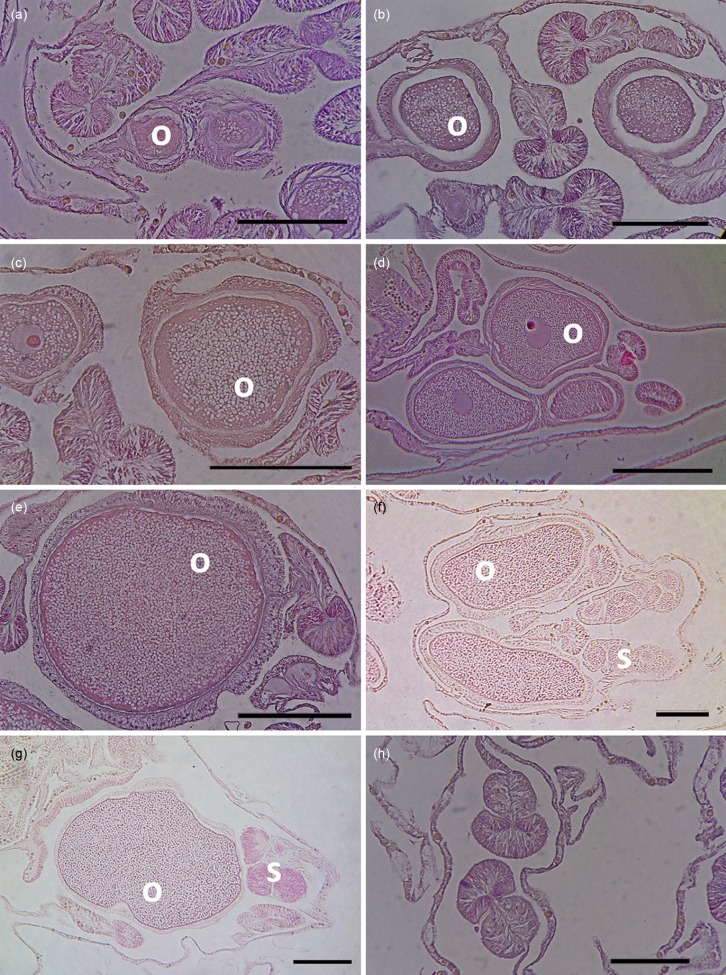
Fig. 3. Histological sections of Acropora hyacinthus in different months: (a) August 2014, (b) October 2014, (c) November 2014, (d) December 2014, (e) January 2015, (f) February 2015, (g) March 2015, (h) April 2015, showing approximate duration of oogenesis of A. hyacinthus at the study location. The oocytes were first observed in some samples in August 2014 and disappeared in all tagged colonies in April 2015. Mature stages of oocytes and spermaries were observed in February and March 2015. o - oocytes; s - spermaries; scale bar = 100 μm.

