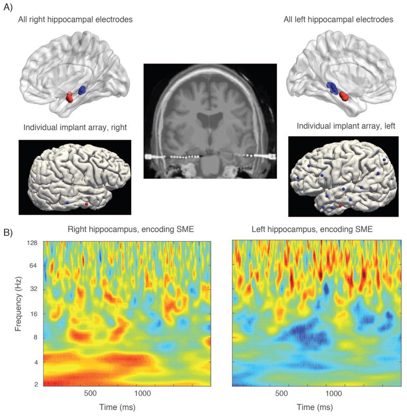
Figure 1. Example of power subsequent memory effect in left and right sided contacts.
A) (top row) shows location of left and right hippocampal contacts across all subjects in the dataset using normalized electrode coordinates. Panels beneath are from a single patient showing electrode locations from a stereo EEG evaluation for a patient contributing anterior and posterior hippocampal data on a surface rendering (electrode entry locations); middle image shows these electrodes in coronal cross section. B) shows the subsequent memory effect in the right and left hippocampus, respectively from the same patient whose electrodes are shown in A. Gamma subsequent memory effect is stronger in the left sided contact for this patient, a pattern that persists across subjects. The right–sided SME also is stronger in the slow–theta range for this subject.

