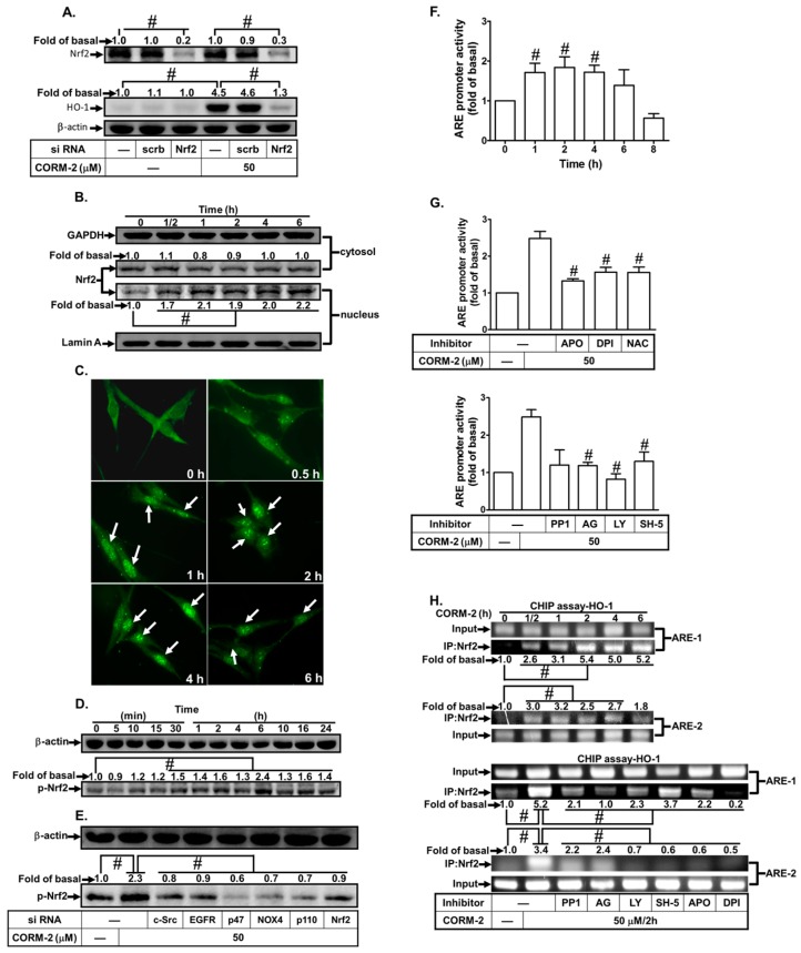
Figure 7.
CORM-2 induces HO-1 expression via Nrf2. (A) HPAEpiCs were transfected with scrambled or Nrf2 siRNA and then incubated with CORM-2 for 16 h. The levels of Nrf2, HO-1, and β-actin protein were determined by western blot. (B) Cells were treated with CORM-2 for the indicated time intervals. The cytosol and nucleus fractions were prepared and subjected to western blot using an anti-Nrf2 antibody. GAPDH and lamin A were used as marker proteins for cytosol and nucleus fractions, respectively. (C) Cells were treated with CORM-2 for the indicated time intervals. Cells were fixed, and then labeled with the anti-Nrf2 antibody and FITC-conjugated secondary antibody. Individual cells were imaged as described in Section 2. (D,E) Cells were treated with CORM-2 for the indicated time intervals or transfected with siRNA of c-Src, EGFR, p47phox, Nox2, or p110, and then incubated with CORM-2 for 6 h. The protein levels of phospho-Nrf2 and β-actin were determined. (F,G) ARE-luc plasmids transfected HPAEpiCs were pretreated (F) without or (G) with PP1, AG1478, LY294002, SH-5, APO, DPI, or NAC for 2 h and then incubated with vehicle or CORM-2 (30 μM) for 2 h. ARE promoter luciferase activity was determined in the cell lysates. (H) Cells were treated with CORM-2 for the indicated times or pretreated with PP1, AG1478, LY294002, SH-5, APO, or DPI, and then incubated with CORM-2 for 6 h. Nrf2 binding activities were analyzed by a ChIP assay. Data are expressed as mean ± S.E.M. of three independent experiments (n = 3). # p < 0.01, as compared with the cells exposed to the CORM-2 alone or compared between the indicated groups.

