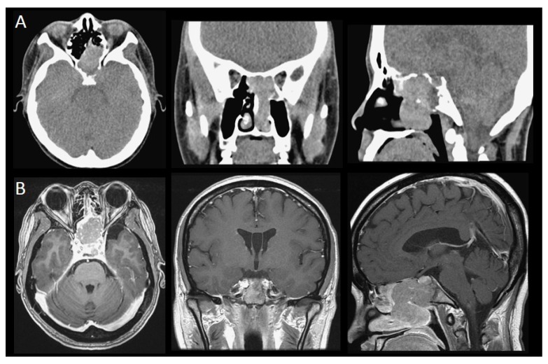
Figure 2.
CT and MRI images of sinonasal metastatic cancers: (A) A 37-year-old female diagnosed with metastatic retroperitoneum leiomyosarcoma. The CT scan revealed that the tumor involved the nasal chamber, bilateral sphenoid sinus, left side of the posterior ethmoid sinus, left aspect of the sellar floor, and the clivus. (B) A 45-year-old female diagnosed with retrorectal neuroendocrine carcinoma. The MRI scans revealed that the tumor involved the sphenoid sinus, sella, suprasella, left cavernous sinus, and pituitary gland.

