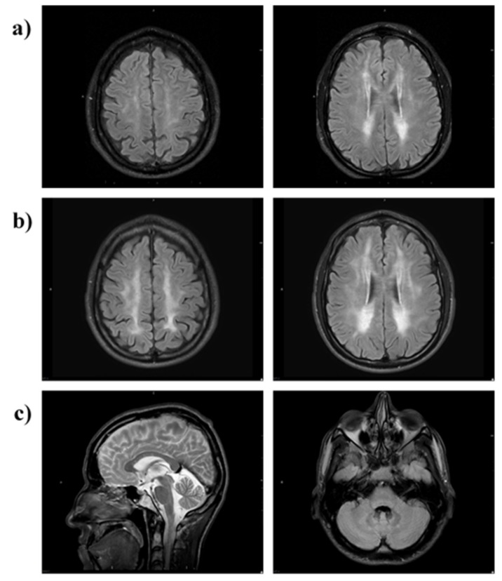
Figure 7.
MRI brain of Patient 2. MRI (axial FLAIR) at (a) 28 months and (b) 63 months shows progression of T2/FLAIR hyperintense cerebral white matter changes (leukoencephalopathy). The scan at 28 months shows patchy involvement of cerebral periventricular, deep and subcortical white matter (a) which 35 months later is more confluent and extensive (b). There is relative sparing of the U-fibres and the corpus callosum (c, left image) and infratentorially there is minor symmetrical FLAIR hyperintensity in the cerebellar peridentate white matter (c, right image).

