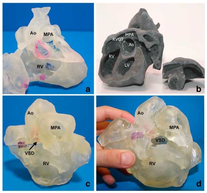
Figure 4.
3D printed heart models showing normal anatomy and pathology. (a) Normal heart model created from cardiac CT and is partitioned into three pieces allowing visualization of interventricular septum. (b) Repaired Tetralogy of Fallot (ToF) from an adult patient. The model was created from cardiac magnetic resonance imaging (MRI) and separated into two pieces allowing for clear visualization of overriding aorta and pulmonary infundibular stenosis. (c) Unrepaired ToF heart model from an infant. The model was created from 3D echocardiographic images and partitioned into two pieces showing the ventricular septal defect (VSD). (d) Unrepaired ToF heart model from an infant with superior and inferior portions showing VSD and the aortic overriding in relation to the VSD. Reprinted with permission under the open access from Loke et al. [22]. Ao—Aorta; MPA—Main Pulmonary Artery; LV—Left Ventricle; RV—Right Ventricle; RVOT—Right Ventricular Outflow Tract; VSD—Ventricular Septal Defect; ToF—Tetralogy of Fallot.

