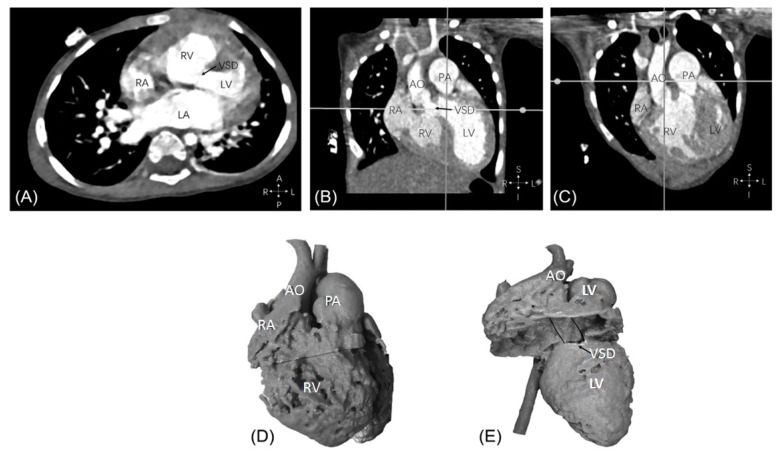
Figure 10.
Example of double outlet right ventricle with aorta and pulmonary artery arising from the right ventricle and perimembranous ventricular septal defect from computed tomography images (A–C). Anterior view of the 3D printed heart model, aorta, and pulmonary artery are side-by-side with both arising from the right ventricle (D). Perimembranous VSD remoted from the arteries. Position of potential intracardiac tunnel from the left ventricle to the aorta is shown as the solid lines (E). AO—ascending aorta; LA—left atrium; LV—left ventricle; PA—pulmonary artery; RA—right atrium; RV—right ventricle; VSD—ventricular septal defect. Reprinted with permission from Zhao et al. [32].

