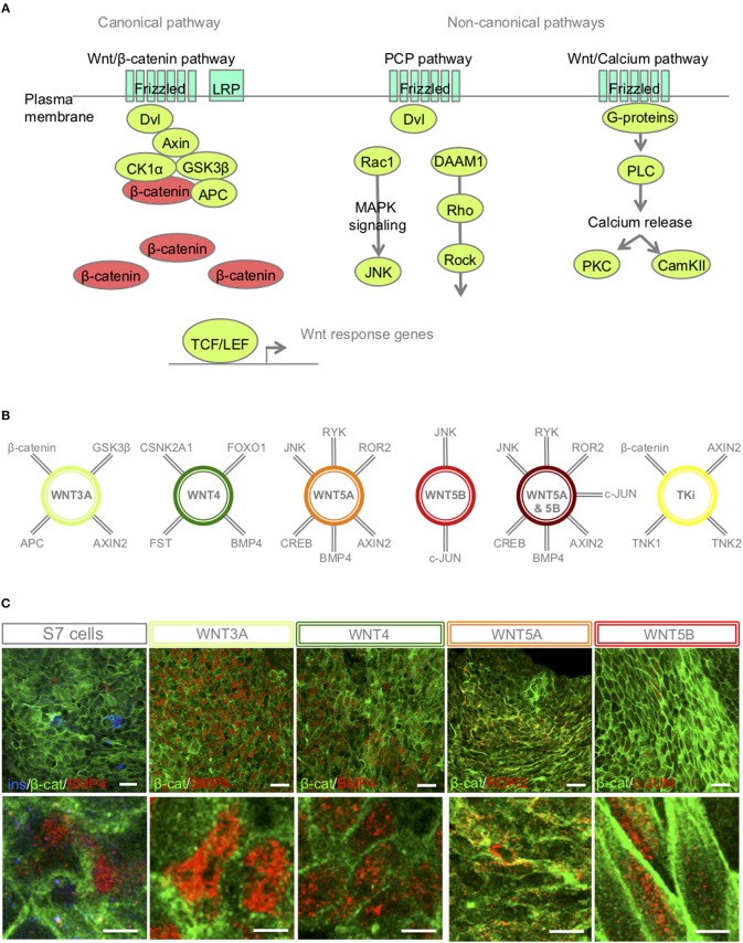
Figure 2.
S7-cells show heterogeneous expression of downstream targets 48 h after Wnt-modulator treatment. (A) Schematic overview of key proteins of the canonical and non-canonical Wnt signaling pathways, in brief, Wnt ligand binds to its receptor Frizzled and co-receptors LRP5/6, receptor tyrosine kinase, (or ROR2) and transmits the signal via disheveled (Dvl) into the cytoplasm to activate the canonical Wnt pathway, or functions through non-canonical planar cell polarity (PCP) and Wnt/Ca2+. In the canonical Wnt signaling pathway, β-catenin accumulates in the cytoplasm and translocate to the nucleus to act as a transcription coactivator for the TCF/LEF transcription factor family. Without Wnt, β-catenin is degraded by the destruction complex, composed of Axin, adenomatosis polyposis coli (APC), glycogen synthase kinase 3β (GSK3β) and casein kinase 1α (CK1α). The non-canonical Wnt/PCP pathway is thought to use Ryk or ROR2 for activation; Dvl is recruited to form a complex with disheveled-associated activator of morphogenesis 1 (DAAM1). DAAM1 activates Rho that again activates Rho-associated kinase (ROCK). Dvl can also form a complex with Rac1 to activate JNK via the MAPK pathway. In the Wnt/Ca2+ pathway, binding of Wnt to Frizzled activates a trimetic G-protein leading to activation of PLC to cleave PIP2 to form DAG and IP3. IP3 binds to its receptor on the endoplasmic reticulum and calcium is released. Increased concentrations of calcium and DAG can again activate PKC and CaMKII. (B) A selection of interaction partners of the selected Wnt-ligands (30–33). (C) IF of β-catenin, BMP4, ROR2 and c-JUN in S7 cells, WNT3A, WNT4, WNT5A, WNT5B treated S7 cells, respectively. Scale bar 50 μm.

