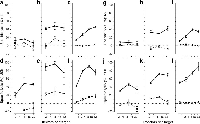
Fig. 2.
Alloreactive and virus-specific CTLs can target hESC-PEs and differentiated hESC-ECs. ESC-PEs (a, b, d, e, g, h, j, k) and hESC-ECs (c, f, i, l) expressing HLA-A1 were labelled with 51Cr and incubated with alloreactive CTLs targeting HLA-A1 (black circles, solid line) or targeting third party HLA-A2 (white circles, dashed line) (a–f) and virus-specific CTLs recognising CMV peptide in HLA-A1 on peptide-pulsed cells (black circles, solid line) or without peptide (white circles, dashed line) (g–l). Specific lysis after 4 h (a–c, g–i) and 20 h (d–f, j–l) was calculated relative to spontaneous lysis without T cells and chemically -induced maximum lysis. Inflammation was mimicked (b, e, h, k) by pre-incubation with IFNγ (1000 IU/ml), which upregulated HLA expression. Statistical results are available in ESM Fig. 1

