
Fig. 1.
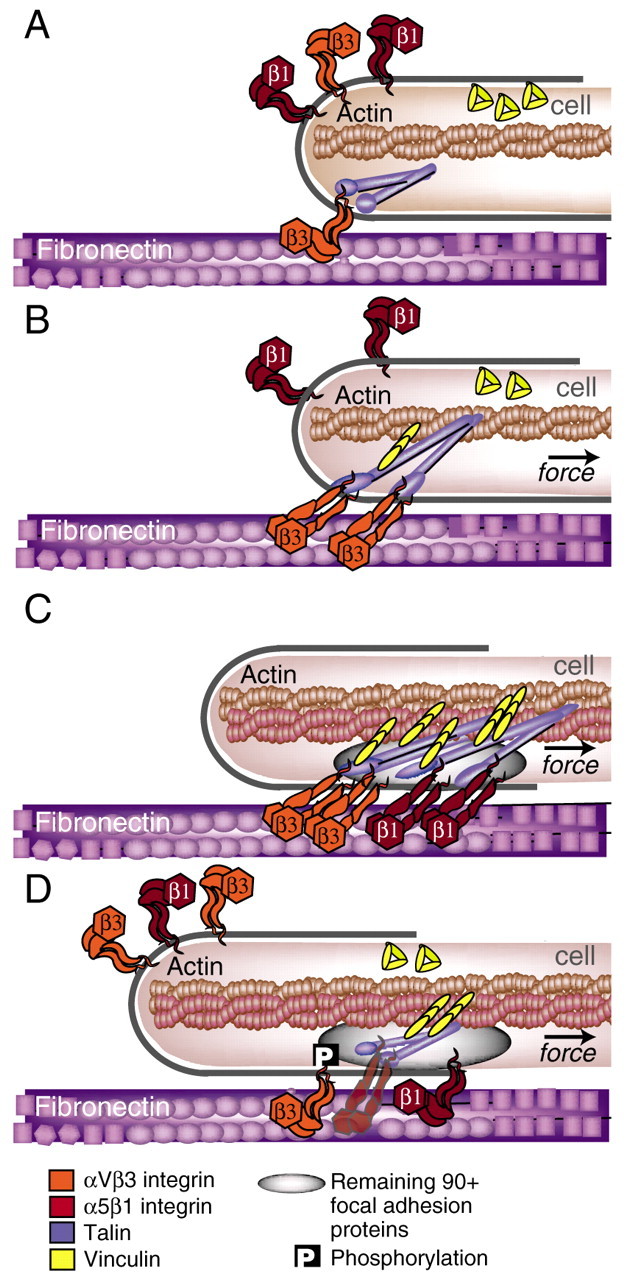
The mechanical integrin cycle. (A) Cell-ECM adhesion occurs when actin-dependent protrusions bring integrins at the leading edge (orange) in contact with the matrix (purple) where they can bind. (B) Next, the integrins link to the actin cytoskeleton through adaptor proteins, such as talin (blue), Shp2, filamin or α-actinin. Integrins bind to these adaptor proteins through their β-tails. Rearward actin flow, generated by actin polymerization and actomyosin contractions (see Box 1) induces a pulling force on the integrin-ECM linkage. On sufficiently rigid substrates, this may serve to accelerate an integrin-activating conformational change, as well as a talin stretch, which may expose buried vinculin-binding sites (yellow). Although the bent conformation of the ligand-bound αVβ3-integrin crystal structure produced much controversy in the integrin field (Liddington and Ginsberg, 2002; Mould et al., 2003), it has subsequently been shown in electron microscopy experiments to stably bind fibronectin (Adair et al., 2005). Force might accelerate the switch to high-binding affinity by freeing the ligand-bound integrin head from the constraints of neighboring domains, which would essentially accelerate the allosteric pathway to the activated state (Puklin-Faucher et al., 2006). (C) The cell begins to pull itself over the site of adhesion. Intramolecular conformational changes in α5β1 integrins facilitate their inward translocation, whereas αVβ3 integrins remain anchored at the edge. This segregation of integrins may further facilitate the talin stretch. At this stage of adhesion, a wide variety of intracellular focal-adhesion proteins are accumulated in the adhesive plaque (grey oval). (D) Ultimately, highly clustered integrins switch from high- to low-binding affinity, possibly catalyzed by the phosphorylation of β3-integrin tails. Membrane exocytosis places recycled, low-affinity integrins at the end of microtubules, often 2-4 μm away from the leading edge. The integrin turnover in focal adhesions (from C to D) is ∼1-3 minutes (Hu et al., 2007). For the description of a single integrin see supplementary material Fig. S1.