Fig. 1.
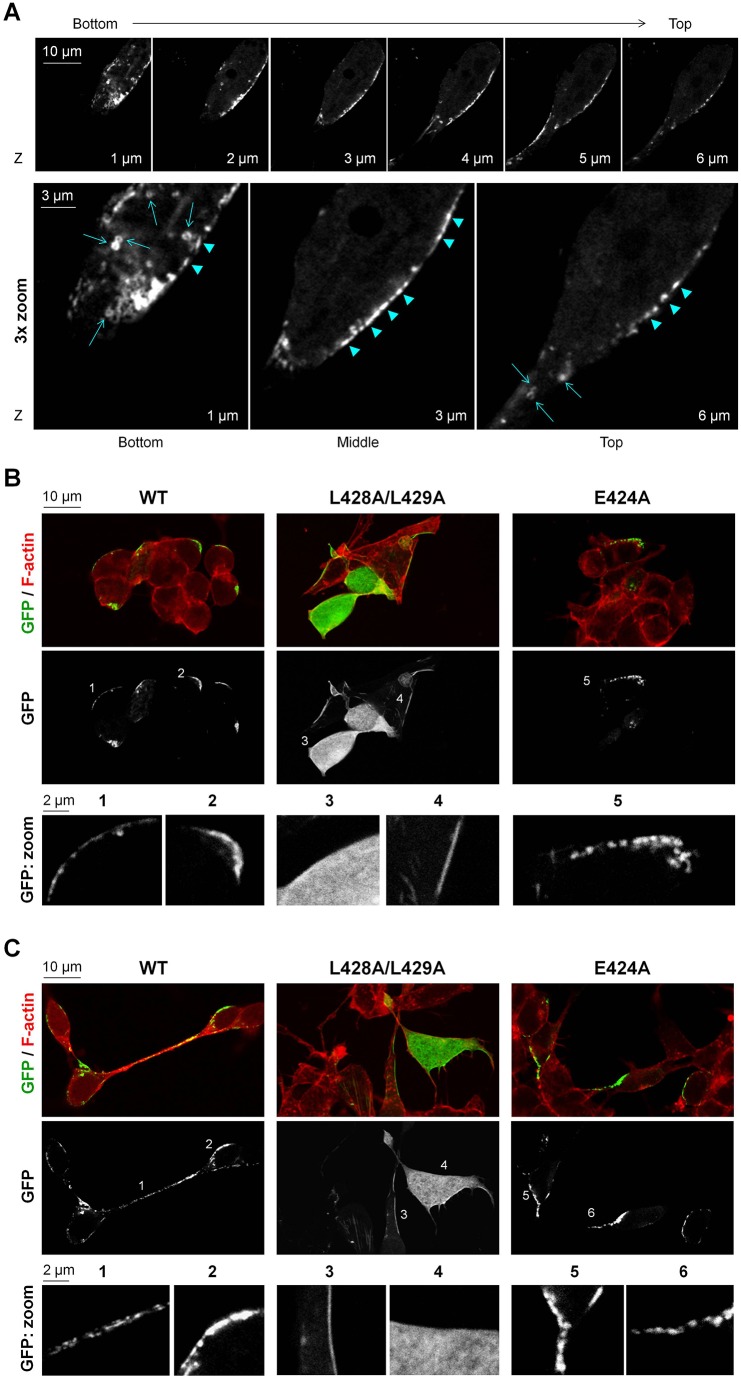
L248A/L249A mutation impairs clustered or small aggregate distribution of LCA at the plasma membrane. (A) Confocal microscopy images showing z-sectioning of a differentiated SiMa cell expressing the wild-type (WT) GFP–LCA. GFP–LCA is seen in ring-like (arrows) and rod-like (arrowheads) clusters or small aggregates at the plasma membrane. The ring-like structures are predominantly seen in the bottom and top sections of the cell, whereas the rod-like structures are mostly seen along the plasma membrane in the middle section of the cell. (B,C) Effect of mutations in the dileucine-based consensus motif on localization of LCA. Confocal microscopy images show loss of clustered or aggregate distribution of LCA owing to the L428A/L429A, but not the E424A, mutation both in non-differentiated (B) and differentiated SiMa cells (C). F-actin (phalloidin) staining was used to show the outlines of cells. The position of the ‘zoom’ images are indicated by numbers.

