
Fig. 2.
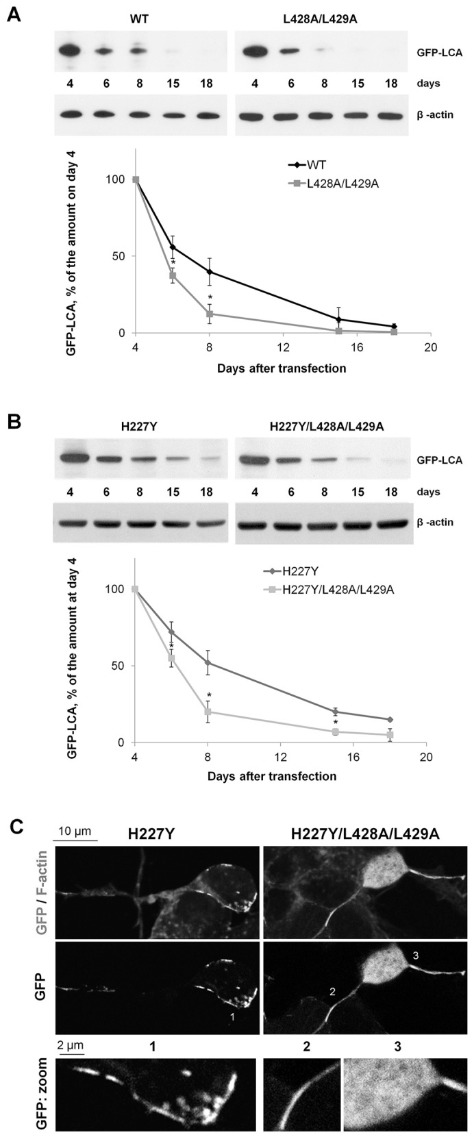
The L428A/L429A mutation decreases the life time of LCA. (A,B) Non-differentiated cells expressing the wild-type (WT) LCA or the L428A/L428A mutant (A) and the H227Y or H227Y/L428A/L428A mutants (B) were lysed after the indicated time periods of cell incubation in low-serum medium following transfection, and the amount of GFP–LCA and β-actin was determined by immunoblotting. Densitometry quantification of the GFP–LCA signal normalized by β-actin signal show that the L428A/L429A mutation shortens the life span of LCA. (C) Confocal microscopy images showing that the H227Y mutation did not change the clustered or aggregate distribution of LCA, whereas the additional mutation of L428 and L429 to alanine residues in the inactive mutant of LCA resulted in a loss of clustered distribution. Non-differentiated SiMa cells incubated for 3 days in a low-serum medium are shown. F-actin (phalloidin) staining was used to show the outlines of cells. Results are mean±s.d. (n = 3). *P<0.01 compared with the wild-type LCA (A) or H227Y (B), Student's t-test.