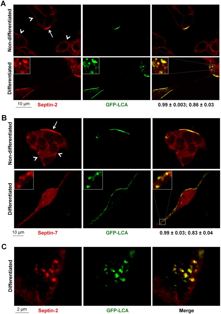
Fig. 4.
LCA recruits septin-2 and septin-7 to membrane-associated clusters or small aggregates. (A,B) LCA is colocalized with septin-2 (A) and septin-7 (B) in clusters at the plasma membrane both in non-differentiated and differentiated SiMa cells. Immunofluorescence of both septin-2 and septin-7 in LCA-positive regions at the plasma membrane (arrows) is much more intense compared to that at the plasma membrane in non-transfected cells (arrowheads). Insets show magnified images of the boxed regions. The coefficient of colocalization with septins (mean±s.d., n = 12) followed by the overlap coefficient (mean±s.d., n = 12) is indicated below the merged images. (C) High-magnification images of the basal sections of non-differentiated LCA-transfected cells showing colocalization of LCA and septin-2 in the ring-like structures.

