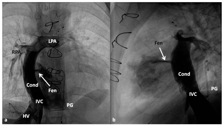
Figure 10.
Selected cine frames in posterio-anterior (a) and lateral (b) views demonstrating Stage IIIA Fontan diverting the inferior vena caval (IVC) flow into the pulmonary arteries via a non-valved conduit (Cond). Flow across the fenestration (Fen) is shown by arrows in (a) and (b). HV, hepatic veins; LPA, left pulmonary artery; PG, pigtail catheter in the descending aorta; RPA, right pulmonary artery. Modified from Rao, P.S. Indian J. Pediatr. 2015, 82, 1147–1156 [79].

