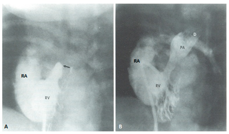
Figure 17.
Selected frames from right ventricular cine-angiograms in a sitting-up view performed prior to (A) and 15 min following (B) perforation of pulmonary valve membrane and balloon pulmonary valvuloplasty. Note the small, heavily trabeculated right ventricle (RV) in (A) without anterograde opacification of the pulmonary artery. The arrow points to the atretic pulmonary valve membrane. Following the procedure, the pulmonary artery (PA) and its branches were well-opacified. Additionally, note opacification of pulmonary end of patent ductus arteriosus (D). There was significant tricuspid insufficiency opacifying the right atrium (RA) both before and after the procedure. Reproduced from Siblini, G.; Rao, P.S.; et al. Cathet. Cardiovasc. Diagn. 1997, 42, 395–402 [24].

