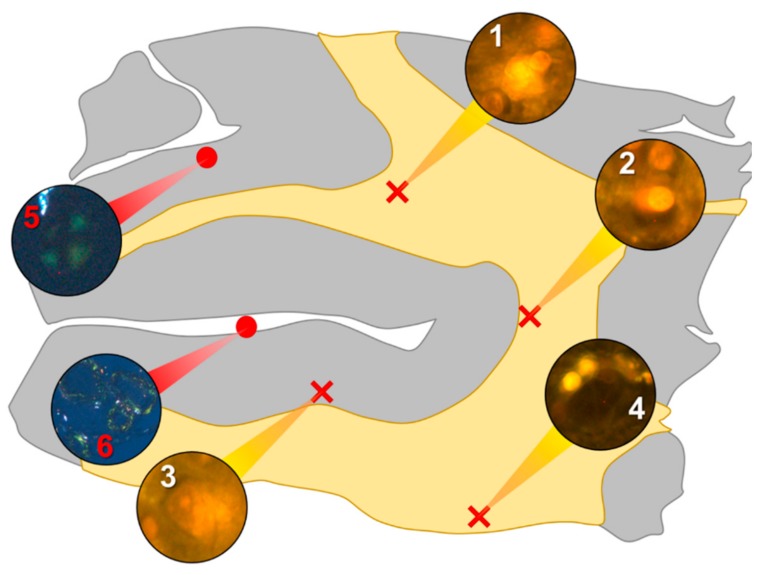
Figure 1.
Schematic depicting a 5 μm tissue section of the parietal lobe. White matter and grey matter are highlighted yellow and grey, respectively. Lumogallion-reactive aluminium was identified through sequential scanning of a 5 μm tissue section, with positive bright orange fluorescence denoted by red crosses (regions 1–4). On an adjacent serial section, Congo red positive regions showing apple-green birefringence under polarised light identified amyloid and spherulites, denoted by red circles (regions 5 and 6).

