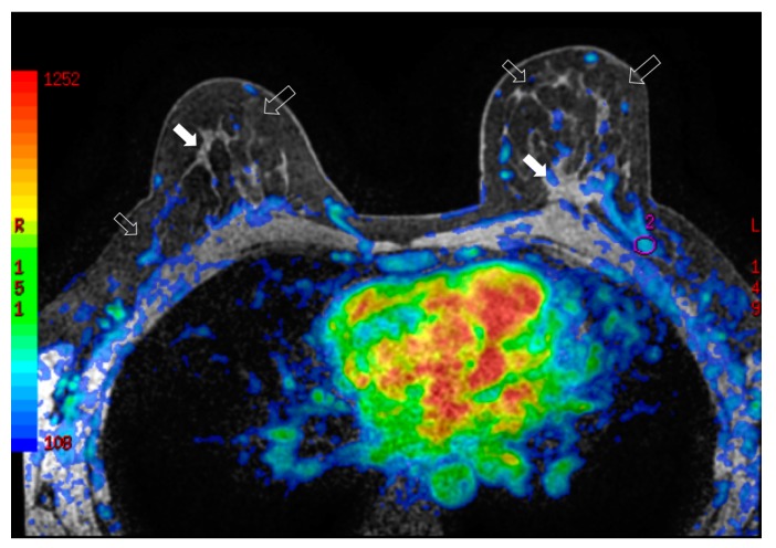
Figure 4.
Bilateral three-dimensional MRI T1-weighted spoiled turbo gradient echo image after contrast media administration (colored superimposed image). Using a special fat saturation and separate shim volumes on each breast, VIBRANT sequence (Volume Imaging for Breast Assessment) allows axial acquisition with an excellent separation of glandular tissue (white arrows) from fat tissue (white empty arrows). The velvet region of interest shows how the vascularity of the left breast compared to the contralateral breast after fat grafting is increased.

