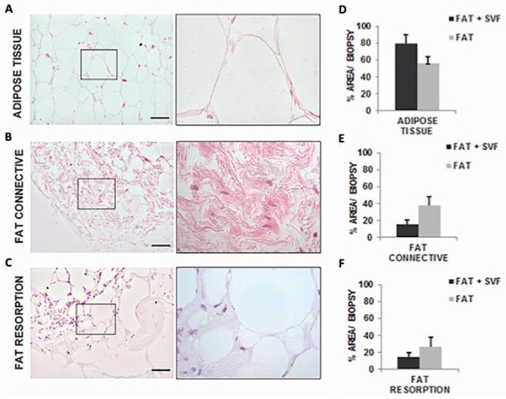
Figure 6.
Images of Hematoxylin and Eosin stained sections (100× magnification and inset 400×) with tissue distribution for the three identified histological features within the biopsies sections. In both groups (SG and CG1) Adipose Tissue (AT) (A) were characterized by round shaped adipocyte without any abnormality. Both groups presented connective tissue (CT) (B), characterized by fibroblastoid cells surrounded by oriented collagen fibers. Fat reabsorption area was an anomalous AT composed by inflammatory cells infiltrating the lobules of adipocytes and associated by small/medium size cysts delimited by polymorphonucleated cells (C). On average, these adipocytes had higher diameter in size versus the normal adipocytic cells. (D–F) showed the comparison of score system results between the two groups in the attempt to investigate the contribution of SVF in the maintenance of AT and CT-reabsorption reduction.

