Fig. 1.
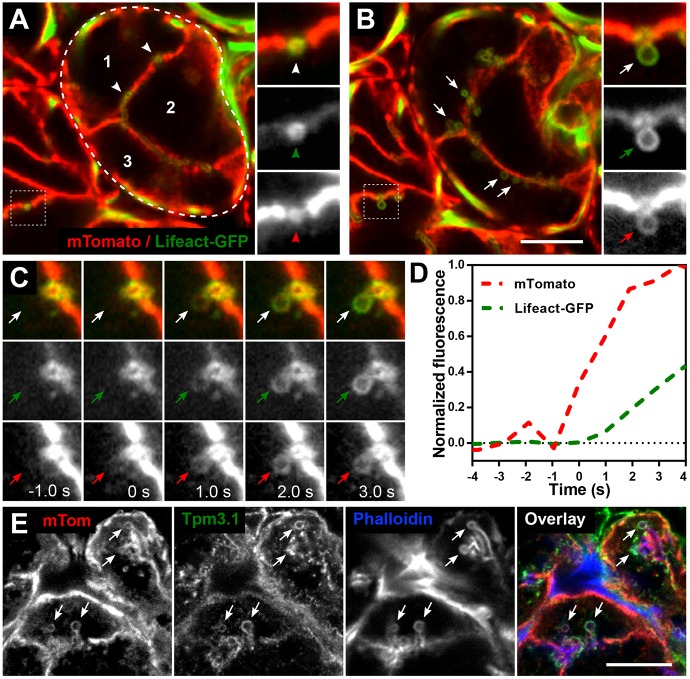
De novo actin and Tpm 3.1 filament polymerisation forms a scaffold around granules after fusion with the APM. (A–D) Intravital confocal imaging of actin filament assembly during secretory granule exocytosis in submandibular salivary gland acinar cells in the progeny of an mTomato and Lifeact–GFP mouse cross. (A,B) Snapshots of salivary acini in situ showing the membrane marker mTomato (red) and F-actin marker Lifeact–GFP (green). (A) Snapshot was taken at a depth of 12 µm from the surface of the gland. Three individual acinar epithelial cells (numbers 1–3) are part of one acinus (encircled by dashed line). Actin is highly enriched at the APM/canaliculi (white arrowheads). Enlarged split channel images displayed on the right show an APM/canaliculus cross-section (red arrowhead) enriched with F-actin (green arrowhead). (B) The same area as in A was imaged 5 min after subcutaneous injection of isoproterenol. Granules fused to APM are seen (white arrows) after stimulation with isoproterenol. Enlarged split channel images show actin recruitment (green arrow) onto the fused granule as seen by the appearance of the mTomato membrane marker (red arrow). Scale bar: 10 µm; the width of the insets is 5.25 µm. See also Fig. S1 and Movie 1. (C) Enlarged time-course images of a representative granule fusion event. The granule acquires an mTomato signal at t=0 s, marking the fusion event (red arrows), and Lifeact–GFP is first detected around the granule circumference at t=1 s (green arrows). Each panel is 5.25 µm wide and the granule diameter is ∼1.2 µm; temporal sampling is 943 ms per frame. (D) Recruitment profiles of mTomato and Lifeact–GFP during a granule fusion event are shown as normalised fluorescence intensity over time. Increase in mTomato (red line) signal from baseline indicates fusion between the granule membrane and the APM. Increase in Lifeact–GFP (green line) indicates actin filament assembly. The frame before the first detection of actin polymerisation was set at t=0 s for this and all subsequent graphs. (E) Detection of mTomato, Tpm3.1 and actin (phalloidin) on fused granules (arrows) in a fixed salivary gland section from an mTomato mouse at 10 min after isoproterenol injection. The contrast was adjusted for each channel separately to facilitate consistent visualisation of the granules in all figures. Scale bar: 10 µm.

