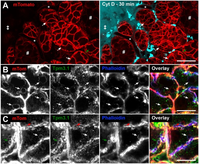
Fig. 2.
Tpm3.1 recruitment onto fused granules is dependent on actin filaments. (A) Overview of intravital imaging of cytochalasin D (CytD) treatment of salivary glands in mTomato mice [stroma (‡), intact capillaries (*), ducts (#)]. Snapshots of glands before (left) and 30 min after (right) CytD treatment. The drug solution contained 10 kDa Alexa Fluor 647 dextran (cyan). CytD treatment for 30 min results in the formation of large vacuoles in acini (white arrows indicate the same acini pre- and post-CytD). CytD penetrance is not uniform, as shown by an unaffected acinus (green arrow) further from the edge of the organ. Enrichment of bright dextran puncta represent rapid dextran marker uptake by the resident stromal cells, such as dendritic cells and fibroblasts. Scale bar: 20 µm. (B,C) Detection of mTomato, Tpm3.1 and actin (phalloidin) on fused granules (arrows) in a salivary gland section of an mTomato mouse treated with CytD (10 min), followed by isoproterenol injection (10 min). (B) CytD-affected granules are enlarged (arrows) and largely devoid of both Tpm3.1 and F-actin staining. (C) The impact of CytD is variable, with some granules exhibiting full (red arrow) or no (green arrow) impact on the actin scaffold. The white arrow shows a fused granule with partial recruitment (crescent-like patch) of F-actin and Tpm3.1. Scale bars: 10 µm.

