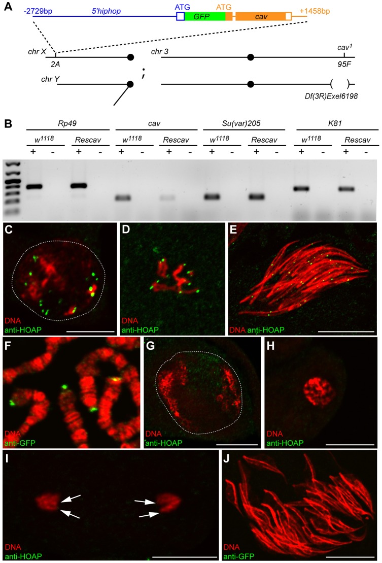
Fig. 3.
cav is downregulated in male germ cells of Rescav flies. (A) A scheme representing the genotype of Rescav males [5′hiphop-GFP::cav/Y; cav1/Df(3R)Exel6198]. The organization of the 5′hiphop-GFP:cav transgene is represented with filled and empty boxes corresponding to, respectively, coding exons and UTRs; and with lines for introns and intergenic regions. The position relative to the ATG of the corresponding gene is indicated. This transgene was inserted into the 2A platform on the X chromosome (chr) and combined with a cav-null allele (cav1) and a deficiency that uncovers cav [Df(3R)Exel6198]. bp, base pair. (B) RT-PCR analyses were performed on total RNAs from the testes of control (w1118) or Rescav flies. Rp49 was used as a control gene. +, with reverse transcriptase; −, without reverse transcriptase. The expression level of cav mRNA is reduced in Rescav testes. The downregulation of cav does not affect the expression of the capping genes K81 and Su(var)205 (encoding HP1a). (C–E) Confocal images of squashed testes from w1118 males stained with an antibody against HOAP (green) and for DNA (red). (C) A nucleus of a primary spermatocyte (the white dashed line delineates the nucleus), (D) metaphase of meiosis II, (E) a cyst of spermatid nuclei. (F) A confocal image of squashed salivary glands of a 5′hiphop-GFP::cav third instar larvae stained with an antibody against GFP (green) and for DNA (red). The fusion protein GFP–HOAP localizes at telomeres of polytene chromosomes. (G–J) Confocal images of squashed testes from Rescav males stained for HOAP or GFP (green) and for DNA (red). (G) A nucleus of a mature primary spermatocyte (white dashed line), (H) prophase of meiosis, (I) anaphase of male meiosis II and (J) nuclei of a cyst of 64 spermatids. In Rescav males, HOAP is undetectable at telomeres (arrows in I) during meiosis and in spermatid nuclei. Scale bars: 10 µm.

