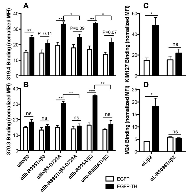
Fig. 6.
Effect of α-integrin CT truncation on talin-1 head induced integrin conformational change. (A,B) Talin-1-head-induced αIIbβ3 epitope exposure for LIBS mAbs 319.4 and 370.3. (C,D) Talin-1-head-induced αLβ2 epitope exposure for mAbs KIM127 and M24. 293FT cells were transfected with the indicated integrin constructs (shown under B and D) and EGFP or EGFP–TH. Cells were first incubated with the biotinylated LIBS mAbs and then stained with PE-labeled streptavidin and Alexa-Fluor-647-labeled mAb AP3 (for αIIbβ3), or stained with Alexa-Fluor-647-labeled streptavidin and PE-labeled TS2/4 (for αLβ2). The EGFP and integrin double-positive cells were analyzed by flow cytometry. The binding of LIBS mAb is presented as the MFI normalized to integrin expression. Data are presented as mean±s.e.m (n≥3). *P<0.05; **P<0.01; ***P<0.001; ns, not significant (unpaired two-tailed Student's t-tests).

