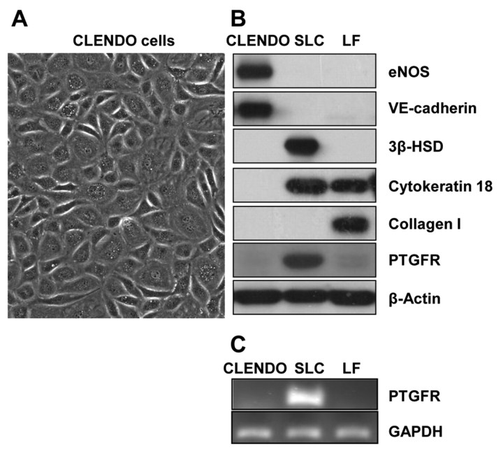
Fig. 1.
Characterization of CLENDO cells. Microvascular endothelial cells were isolated from bovine corpus luteum and characterized by their morphology and expression of cell markers. (A) CLENDO cells displayed cobblestone morphology. CLENDO cells were characterized by western blot of cell lysates (B) and PCR analysis (C). (B) CLENDO expressed the endothelial cell markers VE-cadherin and eNOS, and did not express cytokeratin 18, PTGFR and the specific markers of steroidogenic luteal cells (SLCs) – HSD3B (3β-HSD) – and of luteal fibroblasts (LFs) – collagen I. (C) The receptor for PGF2α, PTGFR, was detected in SLCs, but was not detectable in CLENDO cells or LFs by PCR analysis.

