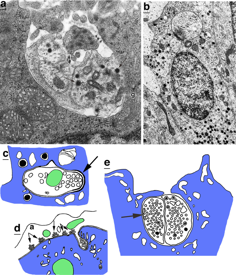
Fig. 12.
Motor nerve endings in endocrine (a-c) and exocrine (d, e) glands. a An axon fascicle is invaginated in a rat adrenal cortical (glomerulosa) cell (g). The lower right process in the fascicle is a synaptic terminal with small, clear vesicles, dense-cored vesicles (arrows) and mitochondria (asterisks). Modified from figure 5B of Kleitman and Holzwarth (1985; reprinted with permission from Springer). b Invaginating terminal (arrow) in an epithelial cell in the pars intermedia of the cat pituitary. Modified from figure 5 in Bargmann et al. (1967; reprinted with permission from Springer). c Drawing of an invaginating terminal in a corticotrope cell of the pars distalis of the dog anterior pituitary. Arrow indicates the thickened postsynaptic density (Ju and Zhang 1990). d Drawing of an axon synapsing on an intercalary cell of the conducting duct of the salivary/silk gland of the mite, Bakericheyla chanayi (Filimonova and Amosova 2015). Note the five active zones (small arrows) formed in a row on the cell surface, followed by an invaginating terminal. e Drawing of an invaginating double terminal in a cell of the labral gland of the water flea, Daphnia obtusa (Zeni and Zaffagni 1988). Arrow, active zone

