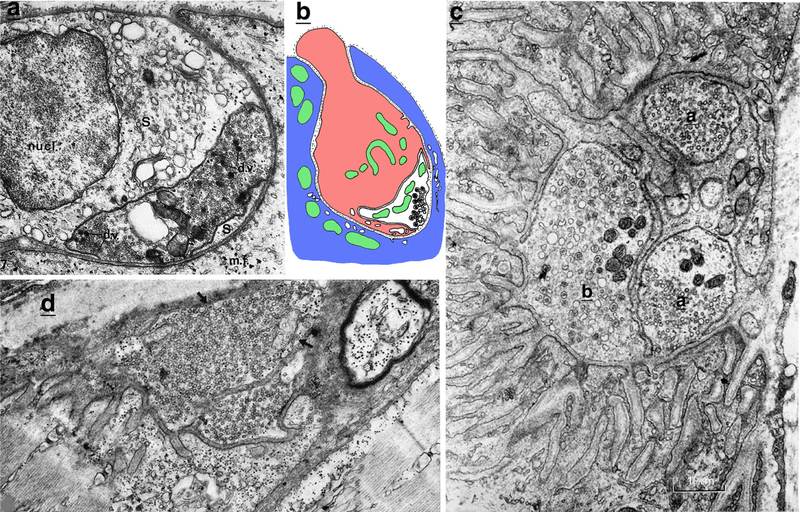
Fig. 9.
NMJs of fish. a, b NMJs of the hagfish, Myxine glutinosa, tend to have indenting/invaginating terminals with Schwann cells that largely fill the top of the invagination. These two examples are from figures of red fibers NMJs in Korneliussen 1973a. a Modified from figure 7 in the latter study (reprinted with permission from Springer); this is an NMJ on a parietal (myotomal) muscle fiber (mf). Note the deep indention that is filled largely by the Schwann cell (S; nucl = nucleus). The presynaptic terminal is at the deep end and so is invaginated completely within the indention; it is filled with small, clear vesicles, as well as some mitochondria and dense-cored vesicles (dv). b Drawing based on figure 9 in the latter study; this is an NMJ on a craniovelar (branchial) muscle fiber. Note how the terminal and Schwann cell (pink) are both enclosed in a deep invagination into the muscle fiber. c The NMJs of dogfish sharks vary; the example shows one of the complex white/twitch fiber NMJs with dual presynaptic terminals (a + b) deeply indenting/invaginating into the muscle fiber (myotomal). Note the extensive subjunctional folds on the left; also, note that some similar surface folds on the right, and extending directly from the muscle fiber surface, are probably myoseptal invaginations (modified from figure 9 in Bone 1972; reprinted with permission from the Company of Biologists Ltd.). Scale bar is 1 micrometer. d Innervation of a myotomal, white muscle fiber close to the myoseptum (upper left of image), from the conger eel, which has better developed subjunctional folds than similar NMJs of most other bony fish (modified from figure 6 in Best and Bone 1973; reprinted with permission from Springer). Arrows show dense-cored vesicles (Color figure online)

