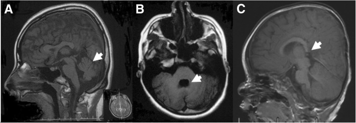
Fig. 1.
Brain abnormality of Patient 1 and Patient 10. (a, b) T1 weighted image of Patient 1 examined at the age of 15 years showed cerebellar vermis dysplasia (a) and fourth ventricle deformity (b). (c) T1 weighted image of Patient 10 examined at the age of 9 months showed dysplasia of the splenium of corpus callosum

