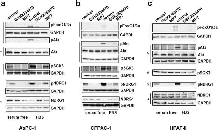
Fig. 4.
Effect of PDK1 pharmacological inhibition on signalling pathways in PDAC cell lines. AsPC-1 (a), CFPAC-1 (b) and HPAF-II (c) cells were serum starved for 24 h and then treated with GSK2334470 (10μΜ) or MP7 (10μΜ) in the presence or absence of FBS for 1h, prior to cell lysis. Lysates were analysed by Western blotting using the indicated antibodies (details of antibodies are provided in the Methods section). In all blots GAPDH was used as loading control. Representative blots are shown. Vertical lines indicate membranes derived from the same gel. In (b,c) membranes incubated with anti-pAkt (Thr308) were stripped and re-incubated with anti-Akt. *, #, $ indicate membranes that derived from the same gel (therefore only one GAPDH is shown)

