Fig. 7.
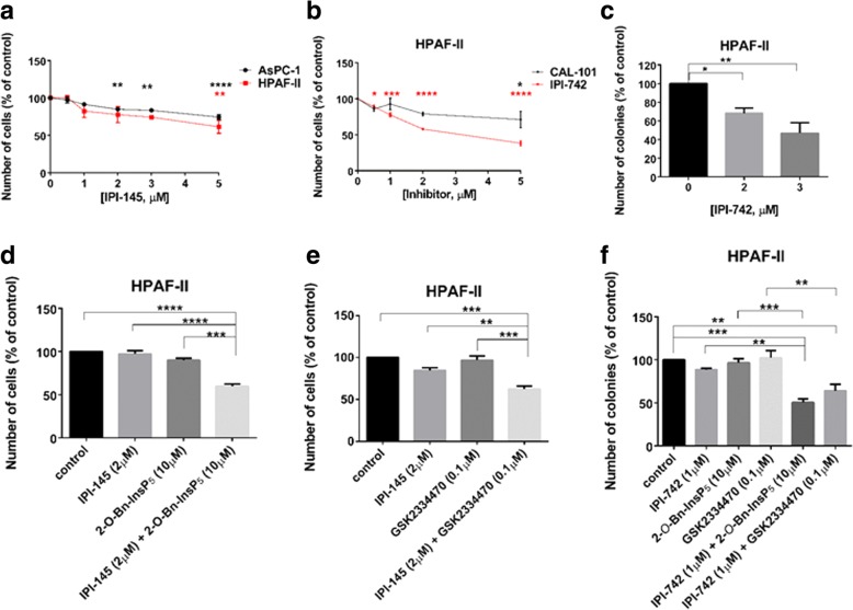
PDK1 inhibitors enhance the effect of p110δ/γ inhibitors on PDAC cell growth. (a,b) HPAF-II and AsPC-1 cells were treated with the indicated concentrations of the p110δ/γ inhibitor IPI-145 (a). Alternatively, HPAF-II cells were treated with the indicated concentrations of the p110δ/γ inhibitor IPI-742 or the selective p110δ inhibitor CAL-101 (b). The number of cells was assessed after 72 h. Data are expressed as percentage of cells treated with vehicle (control) and are means ± SEM of n = 3 independent experiments performed in duplicate. *p < 0.05, **p < 0.01, ***p < 0.001, ****p < 0.0001 vs control cells. (c) HPAF-II cells were plated on soft agar and treated with the indicated concentrations of IPI-742. Data are expressed as percentage of colonies from cells treated with vehicle (control) and are means ± SEM of n = 4 independent experiments performed in duplicate. *p < 0.05, **p < 0.01 vs control. (d, e) HPAF-II cells were treated with IPI-145 (2 μM), 2-O-Bn-InsP5 (10 μM), GSK2334470 (0.1 μM) or the indicated combinations. The number of cells was assessed after 72 h. Data are expressed as percentage of cells treated with vehicle (control) and are means ± SEM of n = 3 independent experiments performed in duplicate. **p < 0.01, ***p < 0.001, ****p < 0.0001. (f) HPAF-II cells were plated on soft agar and treated with IPI-742 (1 μM), 2-O-Bn-InsP5 (10 μM), GSK2334470 (0.1 μM) or the indicated combinations. Data are expressed as percentage of colonies from cells treated with vehicle (control) and are means ± SEM of n = 3 independent experiments performed in duplicate. **p < 0.01, ***p < 0.001

