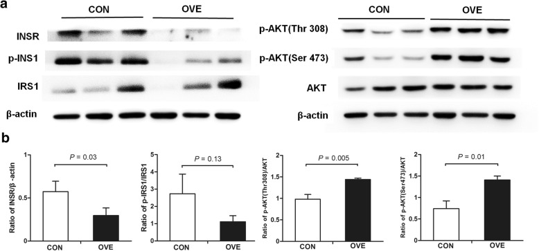
Fig. 4.
Western blot detection of insulin signaling proteins in subcutaneous adipose tissues. These tissues were collected on day 2 postpartum from CON cows (n = 3) and OVE cows (n = 3). a Panels of INSR, IRS1, p-IRS1, AKT, p-AKT (Thr308) and p-AKT (Ser473) protein. β-actin was measured as an internal control. b Intensities of INSR, IRS1, p-IRS1, AKT, p-AKT (Thr308) and p-AKT (Ser473) bands were determined using Quantity One software. The results are presented as the ratio of INSR band intensity to the β-actin band intensity, the ratio of p-ISR1 band intensity to IRS1 band intensity, and the ratio of p-AKT (Thr308) and p-AKT (Ser473) band intensities to the AKT band intensity. IRS1, insulin receptor substrate 1; AKT, protein kinase B

