Fig. 2.
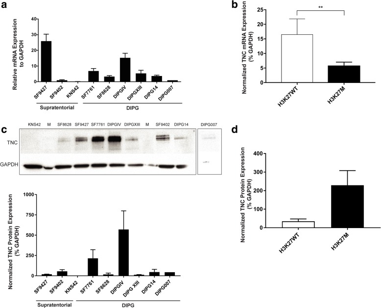
TNC Expression Patterns in Pediatric Glioma Cell Lines. TNC mRNA and protein expression levels were measured in pediatric supratentorial high grade glioma (HGG n = 3) and brainstem glioma (DIPG n = 6) cell lines. a TNC expression determined via quantitative PCR, greatest in DIPGIV (H3.1K27 M) and SF9427 (HGG). b Due to the high level of TNC expressed by SF9427 (H3K27 wild-type), overall TNC mRNA expression was greater in Histone H3K27 wild-type compared to H3K27 M cell lines (**p = 0.0061). a & b Y-axis: Relative TNC mRNA expression level in percent normalized to GAPDH (percent). c TNC protein expression determined via western blot, greatest in DIPGIV and SF7761 (H3.3K27 M mutant lines). d Greater TNC protein expression was seen in Histone H3K27 M compared to wild type cell lines, though this did not reach statistical significance (p = 0.09). c & d Y-axis: TNC protein expression relative to GAPDH (percent). Error bars represent standard error of the mean

