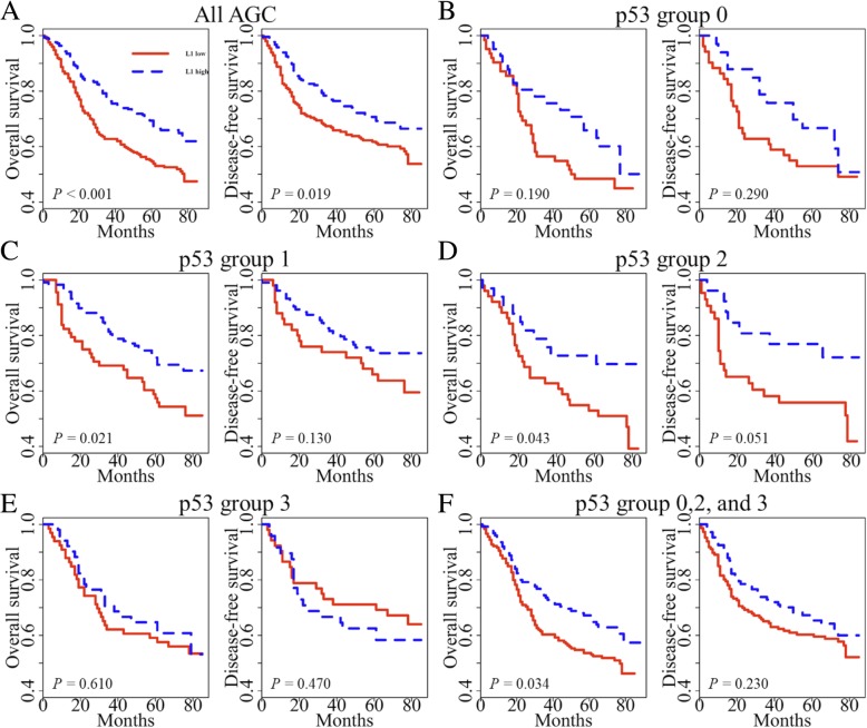
Fig. 3.
Kaplan–Meier survival curves were compared between two groups using a log-rank test to determine difference of patient survival according to L1 methylation status in each p53 expression group. a All AGC samples were included. b AGC samples with negative p53 expression (intensity group 0). c AGC samples with p53 expression group 1. d AGC samples with p53 expression group 2. e AGC samples with p53 expression group 3. f All AGC samples except p53 expression group 1 (group 0, 2, and 3)

