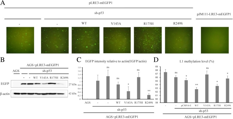
Fig. 4.
L1 expression level and methylation level in TP53-transfected AGS gastric cancer cell line. a Green fluorescence protein observed by fluorescence microscope from TP53-transfected AGS (scale bar 100 μm). b EGFP protein expression levels from TP53-transfected AGS. c Quantification of EGFP intensity by western blot from TP53-transfected AGS. d Mean methylation levels of the four L1 CpG sites for AGS cell line expressing wild type and mutant type TP53 (V143A, R175H, and R249S). (ns P > 0.05, *P < 0.05, **P < 0.01)

