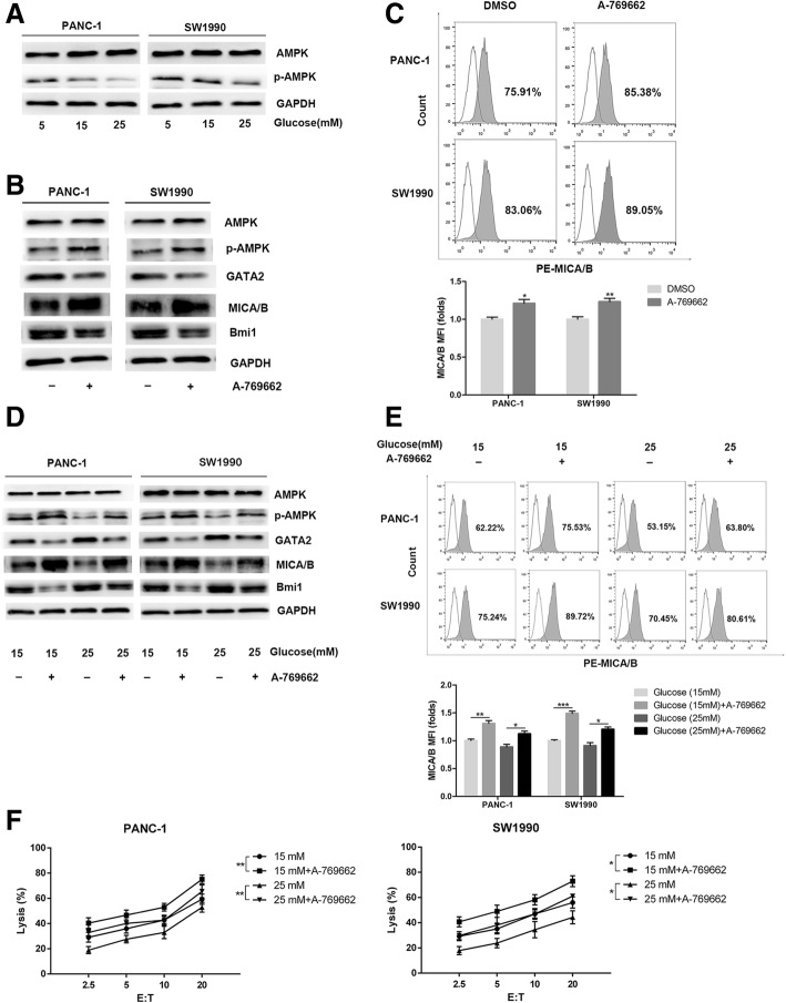
Fig. 6.
High glucose promotes Bmi1 expression through inhibiting AMPK signaling. a Pancreatic cancer cells were treated with different concentrations of glucose for 24 h. Phosphorylation of AMPK was detected with Western blot analysis. b The PANC-1 and SW1990 cells were exposed to the AMPK activator A-769662 (20 μM, 2 h) under normal glucose. The expression levels of Bmi1, GATA2 and MICA/B were detected by Western blot. c Representative histograms of flow cytometry demonstrating MICA/B expression in pancreatic cells treated with AMPK activator. MFI (folds) of MICA/B was evaluated with a Student t test from three independent experiments. d The pancreatic cancer cells were exposed to the AMPK activator A-769662 (20 μM, 2 h) under high glucose. The expression levels of Bmi1, GATA2 and MICA/B were detected by Western blot. e Representative histograms of flow cytometry demonstrating MICA/B expression in pancreatic cells treated with A-69662 under high glucose environment. MFI was evaluated with a Student t test from three independent experiments. f Effect of AMPK activator on the killing ability of NK cells under high glucose. The graphs shown were representative results of three independent experiments. Data in the graphs represented means ± SD from three parallel experiments. **P < 0.01; *P < 0.05

