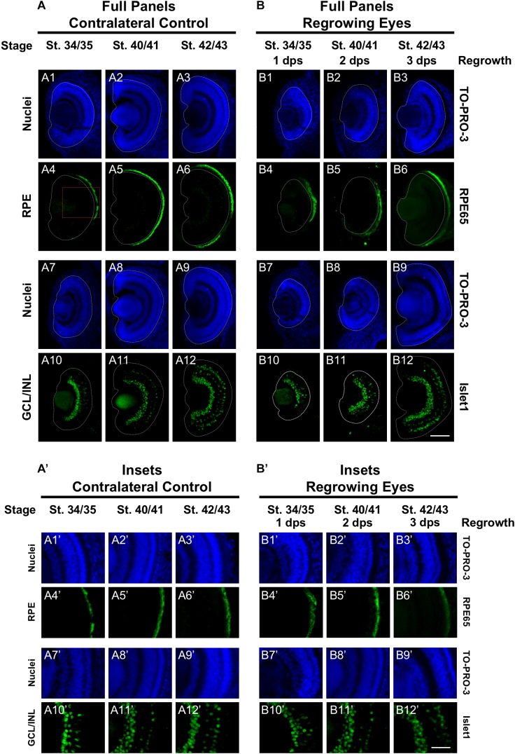
FIGURE 2.
Regrown eyes regain retinal differentiation by 3 dps. Images shown are immunostained, transverse sections at three developmental timepoints corresponding to 1, 2, and 3 days post surgery (dps). (A,B) The contralateral control eyes (unoperated) complete retinogenesis by st. 41. By 1 dps, RPE is already visible in the regrowing eye as shown by anti-RPE65 signal (retinal pigmented epithelium; green). By 3 dps, Islet1 expression (identifying subpopulations of retinal ganglion cells and subsets of amacrine cells, bipolar cells, and horizontal cells; green) show expected retinal patterning of a mature eye. White dashed lines delineate each eye. (A’,B’) Images shown in panels A’ and B’ correspond to the region shown in the inset box in panel A4 for the corresponding (A or B) panel at high magnification. Blue color indicates nuclear staining (TO-PRO-3). Sample sizes: 1 day, n = 5; 2 days, n = 7; and 3 days, n = 6. (A,B, A’,B’) Up = dorsal, down = ventral, lens is on the left. Scale bar: (A,B) = 100 μm and (A’,B’) = 50 μm.

