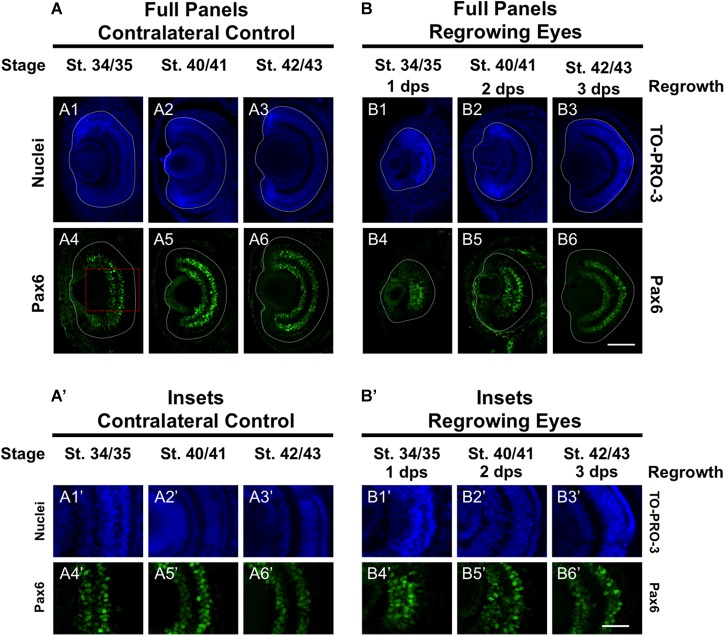
FIGURE 6.
Regrown eyes regain Pax6 patterning by 3 dps. Images shown are immunostained, transverse sections at three developmental timepoints corresponding to 1, 2, and 3 days post surgery (dps). (A,B) Pax6 expression in the regrowing eye is less organized at 1 dps but regains patterning similar to contralateral control eyes (unoperated) by 3 dps. White dashed lines delineate each regrowing eye. (A’,B’) Images shown in panels A’ and B’ correspond to the region shown in the inset box in panel A4 for the corresponding A or B panel at high magnification. Blue color indicates nuclear staining (TO-PRO-3). Green color indicates anti-Pax6 signal. Sample sizes: 1 day, n = 5; 2 days, n = 7; and 3 days, n = 6. (A,B, A’,B’) Up = dorsal, down = ventral, lens is on the left. Scale bar: A,B = 100 μm and A’,B’ = 50 μm.

