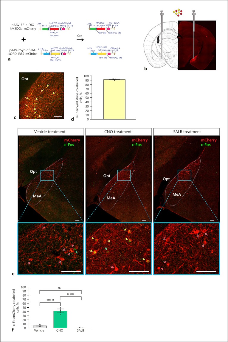
Fig. 3.
Chemogenetic control of MeAKiss neurons in Kiss1CreEGFP/wt male mice. Male Kiss1CreEGFP/wt mice received bilateral MeA stereotaxic injections of an equal mixture of AAV1/2- EF1α-DIO-hM3D(Gq)-mCherry and AAV1/2-hSyn-DIO-hKORD-IRES-mCitrine. After 3 weeks, the mice were treated with CNO (3 µg/g), SALB (10 µg/g), or vehicle (DMSO). a Schematic representation of the AAV vectors encoding hM3D(Gq) and hKORD, both with a FLEX switch allowing recombination (activation) under the control of Cre-recombinase. b Location of stereotaxic viral injection to target Cre-expressing MeAKiss neurons and representative image montage that confirms mCherry expression restricted to the injection site. c Co-localization of hM3D(Gq) (mCherry) and hKORD (mCitrine) marked by yellow arrows. d Quantification of hM3D(Gq) (mCherry) and hKORD (mCitrine) virus co-expression in the MeA (n = 3). e Location of virally expressed hM3D(Gq) (mCherry) and induction of c-Fos (green) 60 min after treatments (vehicle, CNO, or SALB). Yellow arrows mark c-Fos+ virally transduced cells (mCherry), red arrows mark mCherry cells lacking c-Fos expression, and green arrows mark c-Fos+ nuclei. f Quantification of c-Fos+ cells as a percentage of mCherry cells in MeA 60 min after vehicle, CNO, or SALB treatment (n = 3 in each group). Data are represented as mean ± SEM. Circles represent the individual values for each mouse. ns, no significant difference; *** p < 0.001 (one-way ANOVA followed by Tukey's multiple comparison test). opt, optic tract, MeA, medial amygdaloid nucleus. Scale bars, 100 µm.

