Figure 1.
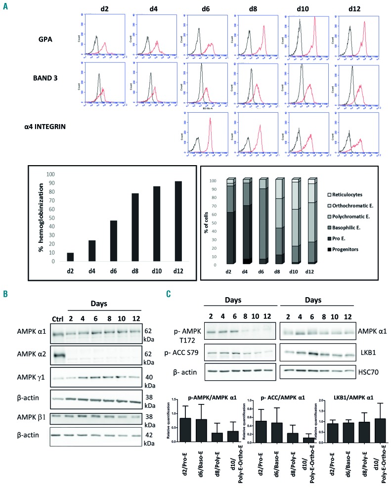
Expression of AMPK isoforms and AMPK activation along terminal erythroid differentiation. (A) Representative experiment of one ex vivo culture of erythroblasts derived from CD34+ cells. Cells were analyzed on days 2, 4, 6, 8, 10 and 12 after CD36+ selection. Expression of cell surface markers GPA, band 3 and α4β1 integrin was studied by flow cytometry along terminal erythroid differentiation. The percentage of hemoglobinized cells was determined by benzidine staining. A minimum of 200 cells were counted and the percentage of blue-stained cells among the total cell count was determined. Cell morphology was examined following staining with May-Grünwald-Giemsa; the percentage of each cell population was determined. (B) AMPK isoforms during erythropoiesis were determined by western blot. Protein extracts from human primary erythroblasts from day 2 to day 12 of culture were analyzed by western blot using anti-α1, -α2, -β1, and -γ1 antibodies. Anti-β-actin was used as a loading control and mouse liver protein extracts were used as a positive control for the expression of AMPKα2 (Ctrl). The α1, α2, and γ1 isoforms were analyzed on the same blot, the β1 isoform on a different one. (C) AMPK activation during erythroid differentiation. Anti-pT172 AMPK, anti-p S79 ACC, anti AMPKα1 and LKB1 were used. Anti-β-actin or anti-HSC70 antibodies were used as loading controls. The upper panel shows a representative experiment of three independent ones. Quantification of western blots and determination of the ratio between pAMPK/AMPK, pACC/AMPK and LKB1/AMPKα1 at the indicated days are presented as the mean of three independent experiments ± SD; ns: non-significant, *P<0.05 (lower panel). d: day; GPA: glycophorin A; AMPK: AMP-activated protein kinase; ACC: acetyl-CoA-carboxylase; LKB1: liver kinase B1; HSC70: heat shock 70kDa protein; E: erythroblasts.

