Figure 2.
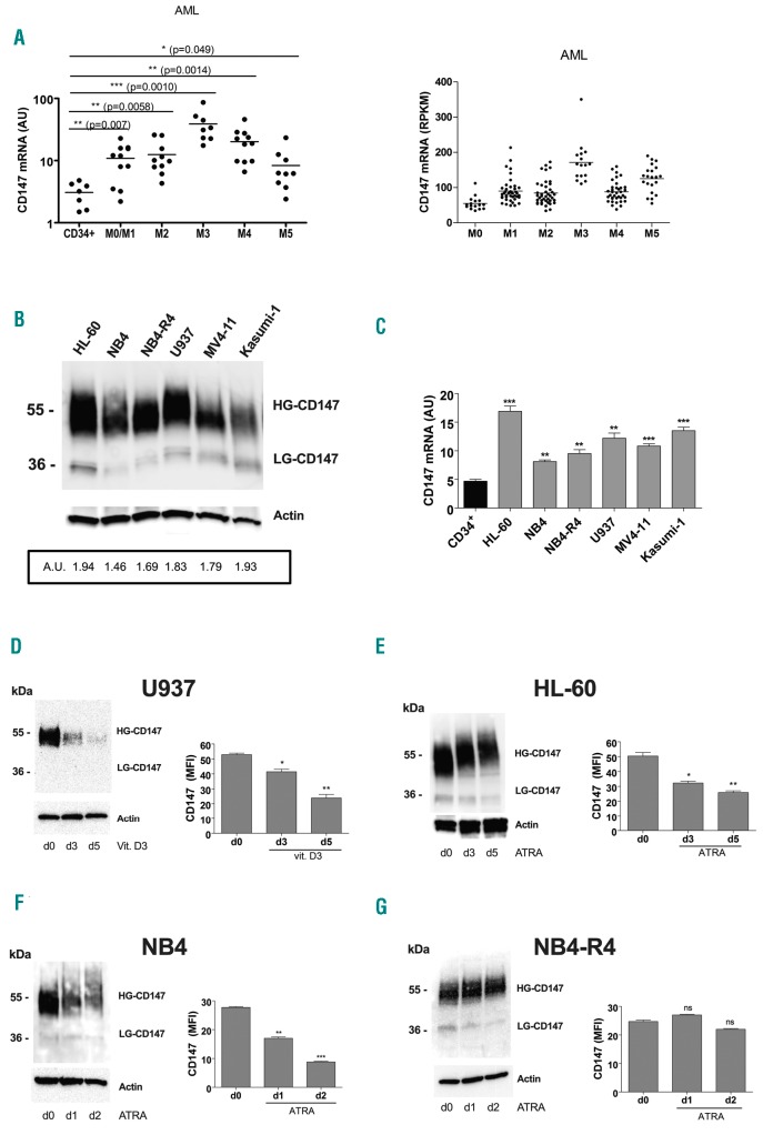
Over-expressed in acute myeloid leukemia (AML), CD147 expression is down-regulated during monocytic and granulocytic differentiation of leukemic cells. (A) qRT-PCR analysis of CD147 mRNA expression in primary leukemic cells of AMLs pertaining from M0 to M5 subtypes of the French-American-British (FAB) classification, as compared to normal CD34+ hematopoietic progenitor cells (HPCs) (left panel); CD147 mRNA expression data from AML samples generated by TCGA Research Network (right panel). (B) Western blot analysis of CD147 protein expression level in AML cell lines; densitometry analysis [arbitrary units (AU)] of CD147 protein expression levels compared with actin levels is indicated. (C) qRT-PCR analysis of CD147 mRNA expression in leukemic cell lines, as compared to normal CD34+ HPCs. (D-G) Western blot (left panels) and flow cytometry (right panels) analysis of CD147 total and membrane protein expression levels during vitamin D3-induced monocytic differentiation of U937 cell (D), ATRA-induced granulocytic differentiation of HL-60 cells (E), ATRA-induced differentiation of NB4 cells (F), and in NB4-R4 cells, resistant to ATRA treatment (G). (A and C) Mean±Standard Error of Mean (SEM) of three independent experiments is shown. *P<0.05; **P<0.01; ***P<0.001. (B, D-G, left panels) One representative western blot experiment out of three is shown. High (HG) and Low (LG) glycosylated CD147 isoforms are indicated. Actin is shown as internal control. (D-G, right panels) Mean±SEM of three independent experiments by flow cytometry analysis is shown. *P<0.05; **P<0.01; ***P<0.001. ns: not significant; RPKM: Reads Per Kilobase Million; MFI: mean fluorescence intensity.

