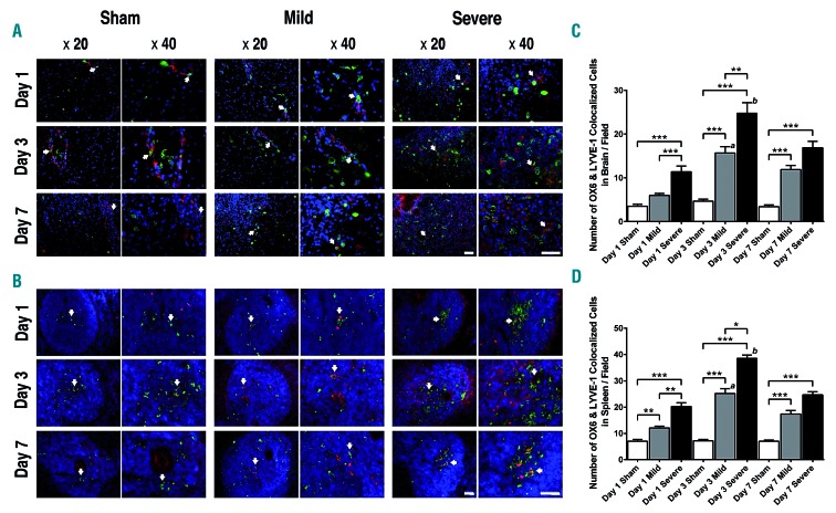
Figure 5.
Localization of OX6-positive cells near or within lymphatic vessels in the brain and spleen. (A and B) Staining with OX6, lymphatic vessel endothelial hyaluronan receptor-1 (LYVE-1), and DAPI, was performed to visualize microglia/macrophages, lymphatic endothelial cells, and nuclei, respectively, in the brain (A) and spleen (B). Arrow heads indicate co-localization of OX6-positive and LYVE-1-positive cells and show that microglia/macrophages localized near or within lymphatic vessels in the brain and spleen. Pictures taken under 20× and 40× magnification. Scale bars = 100 μm. Red: LYVE-1; green: OX6; blue: DAPI. (A) Images were taken close to the dural sinuses in the brain. (B) Images were taken near the gate of the spleen, close to the white pulp in the spleen. (C and D) In quantitative analyses of the brain (C) and the spleen (D), the stroke groups exhibited higher numbers of co-localized cells than did the sham-treated group (**P<0.01). Significance bars: *P<0.05; **P<0.01; ***P<0.001. (C) a: The group with mild stroke had significantly more co-localization in the brain on day 3 than on other days (P<0.05); b: the group with severe stroke had significantly more co-localized cells in the brain on day 3 than on other days (P<0.01). (D) a: The group with mild stroke had significantly more co-localization in the spleen on day 3 than on other days (P<0.05); b: the group with severe stroke had significantly more co-localized cells in the spleen on day 3 than on other days (P<0.01).

