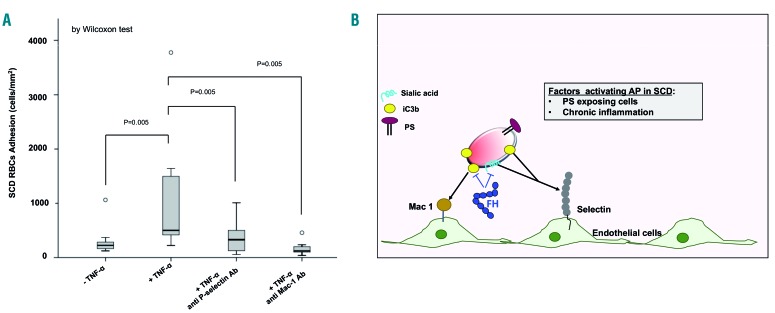
Figure 5.
The anti-adhesive effect of factor H involves P-selectin and Mac-1 pro-adhesive molecules. (A) Adhesion of sickle cell disease red blood cells (SCD RBC) after 6 min of perfusion on activated or non-activated endothelium (± TNF-α) pre-coated with either anti-P-selectin antibody or anti-Mac1 (CD11b/CD18) antibody. Data shown are representative of six other independent assays with similar results. Wilcoxon test: *indicates the corresponding significance. Adhesion is expressed as median values with a minimum-maximum range and illustrated by box plots. A value of P<0.05 is considered statistically significant. Statistical analysis as in Figure 2A. (B) Schematic model of the beneficial action of factor H in reducing adhesion of C3-derived opsonins on sickle RBC to the TNF-α-activated vascular endothelium. C3 split-fragments on erythrocytes might favor cell-cell interactions through P-selectin and Mac-1. P-selectin might bind RBC through two different targets, iC3b and/or sialic acid; in contrast, Mac-1 targets only iC3b deposits on sickle RBC as a more selective interaction. FH and FH19-20 segment normalized the transit of sickle RBC across the TNF-α-activated vascular endothelial surface, abolishing the “stop-and-go” behavior of the sickle RBC. This effect positively affected (shortened) the transit time of sickle RBC, thereby reducing the likelihood of the RBC sickling during their transit through the microcirculation AP: alternative complement pathway; SCD: sickle cell disease; PS: phosphatidylserine; FH: factor H.

