Figure 2.

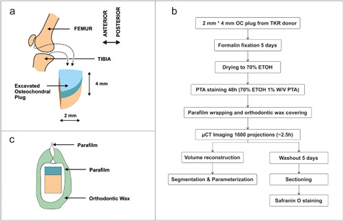
a) Schematics of sample locations of the osteochondral plug. b) Flowchart of OC plug preparation procedure. c) Schematic of sample packaging for μCT imaging.

a) Schematics of sample locations of the osteochondral plug. b) Flowchart of OC plug preparation procedure. c) Schematic of sample packaging for μCT imaging.