FIGURE 1.

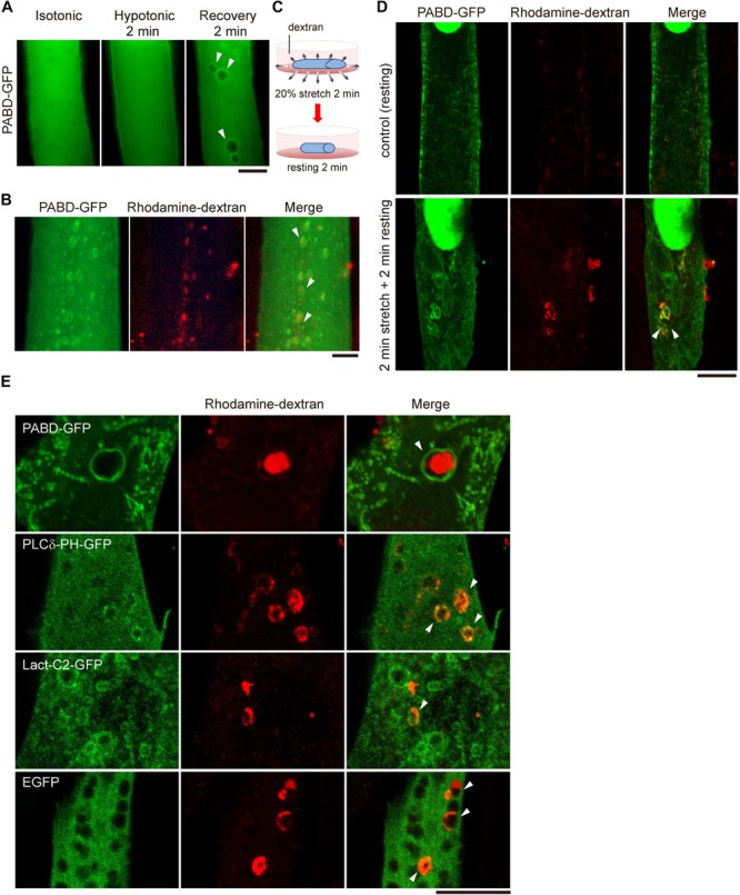
Acute tension fluctuation induces PA-enriched macropinosome formation in myotube. (A) Live cell imaging of PABD-GFP expressing myotube. Myotube was imaged first in isotonic buffer (1X PBS), then in hypotonic buffer (0.25X PBS) upon 2-min incubation, subsequently in recovery isotonic buffer (1X PBS) by 2-min interval. Images of the same myotube were acquired by inverted fluorescence microscope. (B) OS treatment induced the formation of macropinosome. PABD-GFP expressing myotube was treated with OS in the presence of 1 mg/ml rhodamine-dextran. Image was acquired immediately after PBS wash. (C,D) Mechanical stretch induced macropinocytosis in myotube. PABD-GFP expressing myotube cultured on silicon membrane was subjected to 20% radial stretch for 2 min and followed by 2-min resting in the presence of rhodamine-dextran as depicted in (C). After intensive PBS wash, cells were fixed and imaged with confocal microscopy. (E) Distribution of lipid biosensors in myotube upon OS treatment. Myotubes expressing control GFP or biosensors for PA (PABD-GFP), PI(4,5)P2 (PLCδ-PH-GFP) and PS (Lact-C2-GFP) were subjected to OS in the presence of rhodamine-dextran. Projected confocal images were shown. All arrowheads indicate the PA- and rhodamine-dextran containing macropinosomes. Scale bars: 10 μm.
