FIGURE 5.

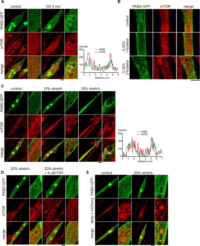
mTOR is localized to the PA-enriched macropinosome. (A) Localization of mTOR in OS-stimulated myotube. PABD-GFP expressing myotubes with or without OS treatment, were immunofluorescence stained to label the endogenous mTOR and were imaged with confocal microscopy. Boxed areas were magnified and shown on the right panel. The fluorescence intensity profiles from the line scan are shown to illustrate the co-localization of mTOR and PABD-GFP. (B) 1-Butanol inhibits the targeting of mTOR to macropinosome upon OS treatment. PABD-GFP expressing myotubes with 0.25% 1- or 2-butanol pre-incubation were subjected to OS treatment and further stained for endogenous mTOR distribution. (C) Single cycle of mechanical stretching and relaxation leads to mTOR targeting to PA-rich macropinosome. PABD-GFP expressing myotubes cultured on silicon membrane were subjected to 0, 10, or 20% radial stretch for 2 min and followed by 2-min resting. After immunofluorescence staining of endogenous mTOR, cells were imaged with confocal microscopy. (D) FIPI inhibits the formation of mTOR- and PA-enriched macropinosome upon stretching. PABD-GFP expressing myotubes were pre-incubated with DMSO or FIPI and then subjected to single cycle of mechanical stretch and relaxation. After fixation and immunofluorescence staining, the distribution of PABD-GFP and endogenous mTOR were imaged with confocal microsocpy. (E) Mechanical stimulation-induced PA is not enriched at lysosome. After single cycle of 20% stretch, PABD-GFP and Lamp1-mCherry expressing myotubes were fixed and imaged with confocal microscopy. Scale bars: 10 μm.
