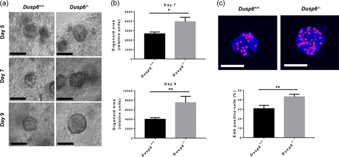
Figure 2.

Dusp6 deletion stimulates colonoid development. (a) Phase‐contrast images of Dusp6 +/+ and Dusp6 ‐/‐ colon organoids after 5, 7, and 9 days of culture. Scale bars: 100 µm. (b) Relative organoid areas after 7 and 9 days of culture (n ≥ 33). (c) EdU incorporation (red) and DAPI staining (blue) were used to evaluate the ratio of proliferative cells per organoid (n = 16). Scale bars: 50 µm. Graphs are representative of at least three independent experiments conducted with different mice. Data are expressed as mean ± SEM. Student‘s t test; *p ≤ 0.05, **p ≤ 0.01. DAPI: 4′,6‐diamidino‐2‐phenylindole; DUSP6: dual‐specificity phosphatase 6; EdU: 5‐ethynyl‐2‐deoxyuridine
