
Figure 3.
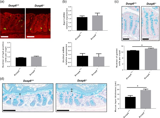
Dusp6 deletion induces goblet cell expansion without affecting enteroendocrine and absorptive cell differentiation. (a) Chromogranin A immunofluorescence with Evan’s blue counterstaining was performed on 12‐week‐old Dusp6 +/+ and Dusp6 −/− murine colon sections to evaluate the number of enteroendocrine cells per crypt. Scale bars=50 µm (n = 7). (b) Relative Ngn3 and Slc26a3 mRNA expression was quantified by qPCR with RNAs isolated from 12‐week‐old murine total colonic extracts (n = 7). Relative expression was normalized to housekeeping genes Pum1, Tbp, and Psmc4 expression. (c) Alcian blue staining was used to visualize Goblet cells in Dusp6 +/+ and Dusp6 ‐/‐ colon sections. Scale bars: 50 µm. The number of Alcian blue positive cells per crypt was counted (n = 7). (d) Carnoy’s fixation followed by Alcian blue staining was performed on 12‐week‐old Dusp6 +/+ and Dusp6 −/− mouse distal colon sections to measure the mucus layer thickness (n ≥ 3). ↕Mucus layer. Scale bars=100 µm. Data are expressed as mean ± SEM. Student’s t test; *p ≤ 0.05. DUSP6: dual‐specificity phosphatase 6; mRNA: messenger RNA; qPCR: quantitaive polymerase chain reaction